PRISM: an energy filtered imaging photoelectron spectrometer
Collaborators:
-
D. Dunham, T. Droubay, J. Denlinger, B. P. Tonner; UW-M
-
Aline Cossy, Javier Diaz, G. Castro, J. Stohr; IBM-Almaden
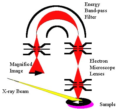
PRISM, which stands for Paraxial Ray Imaging SpectroMicroscope,
is the result of a collaboration between UW-Milwaukee and IBM-Almaden.
It is a new design of a direct-view imaging photoelectron microscope, which
uses electron optics to make a magnified image of a sample surface using
the photoelectrons that are emitted[1]. The electron optics consist of
a high-voltage electrostatic magnification stage, bridged to a low-energy
hemispherical electron spectrometer for selection of the electron kinetic
energy. By tuning the instrument to pass low energy secondary electrons,
which are copious, high spatial resolution images of the surface can be
viewed at standard video rates. To map the surface chemical composition,
the analyzer is set to pass electrons from a specific core-level or valence
band feature, and the image is integrated to build up statistics. A schematic
diagram of the PRISM instrument is shown below.

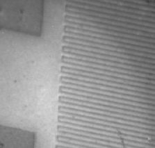
Below is a test image taken using monochromatic light from the BL7 SpectroMicroscopy
Facility undulator beamline. It shows a lithographic pattern in metal on
silicon. The fine lines have a pitch (line-to-line spacing) of 1.2 microns.
Images like this can be acquired at standard video rates.

The most complete reference on PRISM right now is the Ph.D. thesis of
Doug Dunham. Further information is available here.
References:
1 Energy-filtered Imaging with Electrostatic Optics for Photoelectron
Microscopy," B. P. Tonner, Nucl. Inst. and Methods A291, 60-66 (1990).
 Return
to the SpectroMicroscopy Facility home page.
(last updated 27 December 1995 by B. Tonner)


